
Ìtàn ìlò àti ipa àwọn microscopes iṣẹ́-abẹ nínú iṣẹ́-abẹ ọpọlọ
Nínú ìtàn iṣẹ́ abẹ ọpọlọ, lílo tiàwọn microscopes iṣẹ́-abẹjẹ́ àmì àgbàyanu kan, tí ó ń tẹ̀síwájú láti ìgbà ìṣẹ́ abẹ ọpọlọ àtijọ́ ti ṣíṣe iṣẹ́ abẹ lábẹ́ ojú lásán sí ìgbà ìṣẹ́ abẹ ọpọlọ òde òní ti ṣíṣe iṣẹ́ abẹ lábẹ́maikirosikopuTa ni àti ìgbà wo ló ṣe bẹ́ẹ̀àwọn microscopes iṣiṣẹ́bẹ̀rẹ̀ sí í lò ó nínú iṣẹ́ abẹ ọpọlọ?maikirosikopu iṣẹ-abẹṢe a máa ṣe iṣẹ́ abẹ ọpọlọ? Pẹ̀lú ìlọsíwájú ìmọ̀ sáyẹ́ǹsì àti ìmọ̀ ẹ̀rọ, ṣé a máa ṣe bẹ́ẹ̀?Maikirosikopu iṣiṣẹṢé a lè fi àwọn ohun èlò tó ti pẹ́ jù rọ́pò wọn? Ìbéèrè yìí ni gbogbo oníṣẹ́ abẹ ọpọlọ gbọ́dọ̀ mọ̀ nípa rẹ̀ kí ó sì lo ìmọ̀ ẹ̀rọ àti ohun èlò tuntun fún iṣẹ́ abẹ ọpọlọ, èyí tó ń mú kí òye iṣẹ́ abẹ ọpọlọ sunwọ̀n sí i.
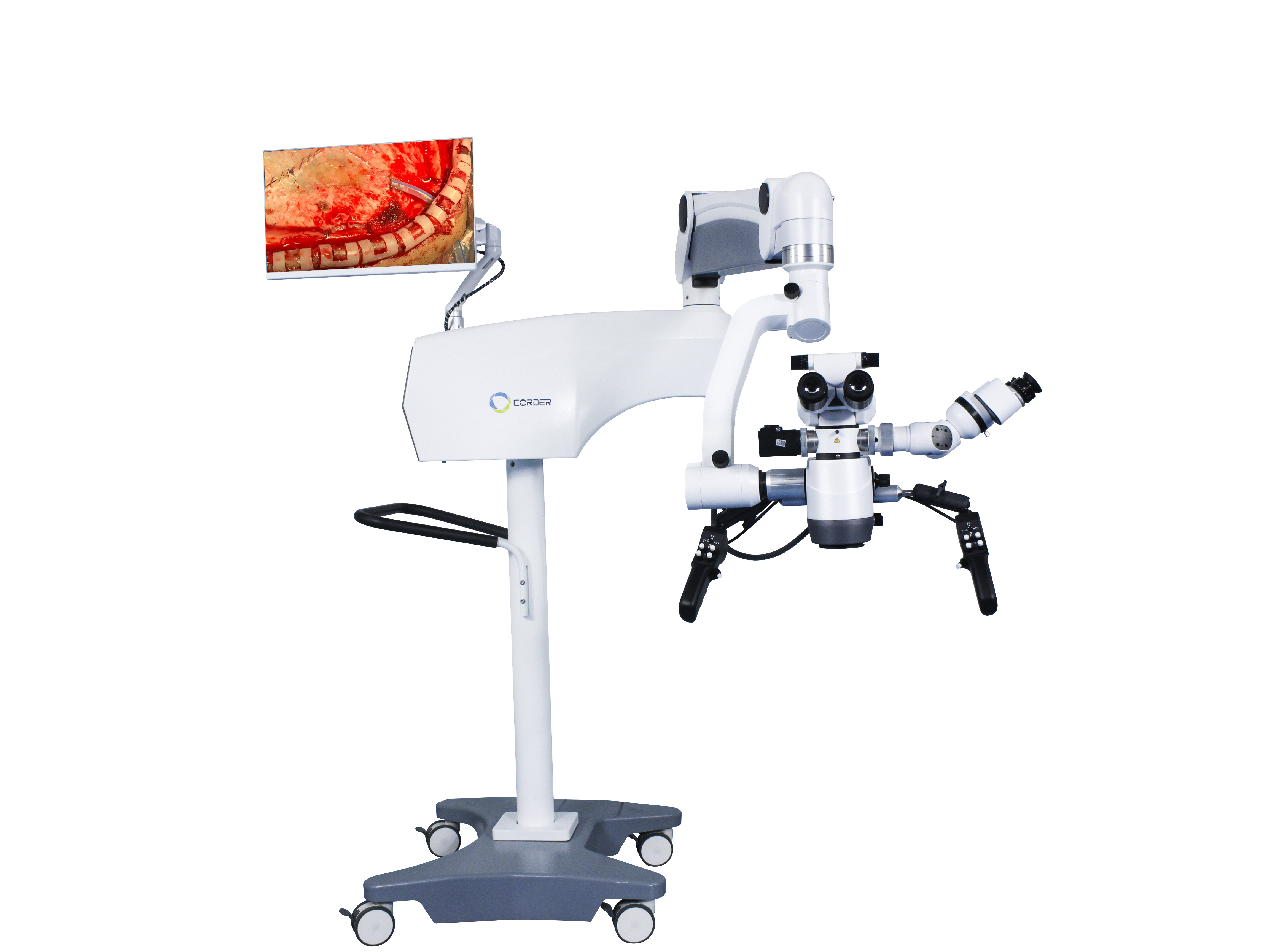
1, Ìtàn Àwọn Ohun Èlò Onímọ̀-ẹ̀rọ-afẹ́fẹ́ ní Iṣẹ́ Ìṣègùn
Nínú ìmọ̀-ẹ̀rọ fisiksi, àwọn lẹ́nsì ojú jẹ́ lẹ́nsì convex pẹ̀lú ìṣètò kan ṣoṣo tí ó ní ipa ìgbéga, àti ìgbéga wọn ní ààlà, tí a mọ̀ sí àwọn lẹ́nsì ìgbéga. Ní ọdún 1590, àwọn ènìyàn Dutch méjì fi àwọn àwo lẹ́nsì convex méjì sínú àgbá onígun mẹ́rin kan tí ó tẹ́ẹ́rẹ́, nípa bẹ́ẹ̀ wọ́n ṣe àwárí ẹ̀rọ ìgbéga ìṣètò àkọ́kọ́ ní àgbáyé:maikirosikopuLẹ́yìn náà, a máa ń mú kí ìṣètò ohun tí a fi ń ṣe microscope náà sunwọ̀n sí i nígbà gbogbo, ìtóbi rẹ̀ sì ń pọ̀ sí i nígbà gbogbo. Ní àkókò yẹn, àwọn onímọ̀ sáyẹ́ǹsì ló sábà máa ń lo èyímaikirosikopu akojọpọláti kíyèsí àwọn ìrísí kékeré ti àwọn ẹranko àti ewéko, bí ìrísí àwọn sẹ́ẹ̀lì. Láti àárín sí ìparí ọ̀rúndún 19, àwọn awòran gíga àti àwọn microscopes ni a ti lò díẹ̀díẹ̀ nínú iṣẹ́ ìṣègùn. Ní àkọ́kọ́, àwọn oníṣẹ́ abẹ lo àwọn awòran gíga bíi gilasi pẹ̀lú ìrísí lẹ́ǹsì kan ṣoṣo tí a lè gbé sórí afárá imú fún iṣẹ́ abẹ. Ní ọdún 1876, dókítà ará Germany Saemisch ṣe iṣẹ́ abẹ "àwòran gíga" àkọ́kọ́ ní àgbáyé nípa lílo awòran gíga dígísáàtì (a kò mọ irú iṣẹ́ abẹ náà). Ní ọdún 1893, ilé-iṣẹ́ ará Germany Zeiss ṣe àgbékalẹ̀ rẹ̀maikirosikopu oju-ọna meji, tí a sábà máa ń lò fún àkíyèsí àyẹ̀wò ní àwọn ilé ìwòsàn ìṣègùn, àti fún àkíyèsí àwọn ọgbẹ́ inú àti iwájú yàrá ní ẹ̀ka ìṣègùn ojú. Ní ọdún 1921, tí a gbé kalẹ̀ lórí ìwádìí yàrá lórí anatomi etí inú ẹranko, onímọ̀ nípa otolaryngologist ará Sweden, Nylen, lo ohun èlò tí a ti ṣe àtúnṣe.microscope iṣẹ-abẹ monocularṢe apẹrẹ ati ṣe ni ara rẹ lati ṣe iṣẹ abẹ otitis media onibaje lori eniyan, eyiti o jẹ iṣẹ abẹ microscopic gidi. Ni ọdun kan lẹhinna, dokita giga Nylen Hlolmgren ṣe agbekalẹ kanmicroscope iṣẹ-abẹ binocularZeiss ṣelọpọ ni yara iṣẹ abẹ.
Àwọn ìbẹ̀rẹ̀Àwọn microscopes iṣiṣẹ́ní ọ̀pọ̀lọpọ̀ àléébù, bí àìdúróṣinṣin ẹ̀rọ tí kò dára, àìlègbéra, ìmọ́lẹ̀ àwọn àáké tó yàtọ̀ síra àti gbígbóná lẹ́ńsì ohun tí a lè fojú rí, pápá ìṣe-abẹ tóóró, àti bẹ́ẹ̀ bẹ́ẹ̀ lọ. Gbogbo àwọn ìdí wọ̀nyí ló ń dín lílo gbogbogbòò kùàwọn microscopes iṣẹ́-abẹNí ọgbọ̀n ọdún tó tẹ̀lé e, nítorí ìbáṣepọ̀ rere láàárín àwọn oníṣẹ́ abẹ àtiawọn oluṣe microscope, iṣẹ́ tiàwọn microscopes iṣẹ́-abẹa n mu ilọsiwaju wa nigbagbogbo, atiàwọn ohun èlò ìṣẹ́ abẹ abẹ́ ojú ìwòran, àwọn microscopes tí a gbé sórí òrùlé, awọn lẹnsi zoom, imọlẹ orisun ina coaxial, awọn apa ti a fi agbara mu nipasẹ ẹrọ itanna tabi titẹ omi, iṣakoso pedal ẹsẹ, ati bẹẹbẹ lọ ni a ṣe agbekalẹ ni atẹlera. Ni ọdun 1953, ile-iṣẹ German Zeiss ṣe agbejade awọn eto pataki kan.àwọn microscopes iṣẹ́-abẹ fún ìmọ̀ nípa otology, o dara julọ fun awọn iṣẹ abẹ lori awọn ọgbẹ jinle bi eti arin ati egungun akoko.àwọn microscopes iṣẹ́-abẹBí àpẹẹrẹ, àwọn dókítà ará Jámánì Zollner àti Wullstein sọ pé, èrò àwọn oníṣẹ́ abẹ náà ń yí padà nígbà gbogbo.àwọn microscopes iṣẹ́-abẹa gbọ́dọ̀ lò ó fún iṣẹ́ abẹ ìrísí awọ ara tympanic. Láti ọdún 1950, àwọn onímọ̀ nípa ojú ti yí àṣà lílo àwọn ohun èlò amúṣẹ́jú nìkan padà díẹ̀díẹ̀ fún àwọn àyẹ̀wò ojú, wọ́n sì ti ṣe àgbékalẹ̀ rẹ̀àwọn ohun èlò amúṣẹ́-abẹ otosurgicalsinu iṣẹ abẹ oju. Lati igba naa,Maikirosikopu iṣiṣẹti lo ni ibigbogbo ni awọn aaye ti opolo ati oju.
2. Lilo microscopes abẹ ninu iṣẹ abẹ ọpọlọ
Nítorí pé iṣẹ́ abẹ ọpọlọ ṣe pàtàkì, lílo rẹ̀àwọn microscopes iṣẹ́-abẹ nínú iṣẹ́-abẹ ọpọlọÓ pẹ́ díẹ̀ ju ti ìmọ̀ nípa ojú àti ìtọ́jú ojú lọ, àwọn oníṣẹ́ abẹ ọpọlọ sì ń kọ́ ìmọ̀ ẹ̀rọ tuntun yìí ní kíákíá. Ní àkókò yẹn,lilo awọn microscopes iṣẹ-abẹÓ wà ní Yúróòpù jùlọ. Onímọ̀ nípa ojú ará Amẹ́ríkà Perrit ló kọ́kọ́ ṣe àgbékalẹ̀ rẹ̀àwọn microscopes iṣẹ́-abẹláti Yúróòpù sí Amẹ́ríkà ní ọdún 1946, ó fi ìpìlẹ̀ lélẹ̀ fún àwọn oníṣẹ́ abẹ ọpọlọ Amẹ́ríkà láti lòÀwọn microscopes iṣiṣẹ́.
Láti ojú ìwòye bíbọ̀wọ̀ fún iye ẹ̀mí ènìyàn, èyíkéyìí ìmọ̀ ẹ̀rọ tuntun, ohun èlò, tàbí ohun èlò tí a lò fún ara ènìyàn yẹ kí ó ṣe àwọn àyẹ̀wò ẹranko àkọ́kọ́ àti ìdánilẹ́kọ̀ọ́ ìmọ̀ ẹ̀rọ fún àwọn oníṣẹ́. Ní ọdún 1955, oníṣẹ́ abẹ ọpọlọ ará Amẹ́ríkà Malis ṣe iṣẹ́ abẹ ọpọlọ fún àwọn ẹranko nípa lílo abẹ́rẹ́ kanmicroscope iṣẹ-abẹ binocularKurze, oníṣẹ́ abẹ ọpọlọ ní Yunifásítì Gúúsù California ní Orílẹ̀-èdè Amẹ́ríkà, lo ọdún kan ní kíkọ́ àwọn ọ̀nà iṣẹ́ abẹ nípa lílo microscope nínú yàrá ìwádìí lẹ́yìn tí ó wo iṣẹ́ abẹ etí lábẹ́ microscope. Ní oṣù kẹjọ ọdún 1957, ó ṣe iṣẹ́ abẹ neuroma acoustic lórí ọmọ ọdún márùn-ún kan ní àṣeyọrí nípa lílo ohun èlò ìṣiṣẹ́ acousticmaikirosikopu iṣẹ abẹ etí, èyí tí ó jẹ́ iṣẹ́ abẹ kékeré àkọ́kọ́ ní àgbáyé. Láìpẹ́ lẹ́yìn náà, Kurze ṣe àṣeyọrí lórí ìṣàn iṣan ara ojú ọmọ náà nípa lílo ohun èlò kan tí a fi ń ṣe iṣẹ́ abẹ kékeré.maikirosikopu iṣẹ-abẹ, àti pé ìlera ọmọ náà dára gan-an. Iṣẹ́ abẹ kékeré kejì ni èyí ní àgbáyé. Lẹ́yìn náà, Kurze lo àwọn ọkọ̀ akẹ́rù láti gbéÀwọn microscopes iṣiṣẹ́si awọn ibi oriṣiriṣi fun iṣẹ abẹ ọpọlọ kekere, ati pe o gbani nimọran gidigidi lati loàwọn microscopes iṣẹ́-abẹfún àwọn oníṣẹ́ abẹ ọpọlọ mìíràn. Lẹ́yìn náà, Kurze ṣe iṣẹ́ abẹ ìdènà àrùn ọpọlọ nípa lílo ohun kanmaikirosikopu iṣẹ-abẹ(ó bani nínú jẹ́ pé kò tẹ àwọn àpilẹ̀kọ kankan jáde). Pẹ̀lú ìtìlẹ́yìn aláìsàn trigeminal neuralgia kan tí ó tọ́jú, ó dá yàrá ìwádìí neurosurgery microskull base àkọ́kọ́ ní àgbáyé sílẹ̀ ní ọdún 1961. A gbọ́dọ̀ máa rántí àfikún Kurze sí microsurgery nígbà gbogbo kí a sì kọ́ ẹ̀kọ́ láti inú ìgboyà rẹ̀ láti gba àwọn ìmọ̀ ẹ̀rọ àti èrò tuntun. Síbẹ̀síbẹ̀, títí di ìbẹ̀rẹ̀ ọdún 1990, àwọn oníṣẹ́ abẹ ọpọlọ kan ní China kò gbà.Àwọn microscopes iṣẹ́-abẹ ọpọlọfún iṣẹ́-abẹ. Èyí kì í ṣe ìṣòro pẹ̀lúMaikirosikopu iṣẹ-abẹ ọpọlọfúnra rẹ̀, ṣùgbọ́n ìṣòro pẹ̀lú òye èrò àwọn oníṣẹ́ abẹ ọpọlọ.
Ní ọdún 1958, oníṣẹ́ abẹ ọpọlọ ọmọ ilẹ̀ Amẹ́ríkà, Donaghy, dá ilé ìwádìí àti ìdánilẹ́kọ̀ọ́ iṣẹ́ abẹ kékeré àkọ́kọ́ sílẹ̀ ní Burlington, Vermont. Ní ìbẹ̀rẹ̀, ó tún rí ìdàrúdàpọ̀ àti ìṣòro owó láti ọ̀dọ̀ àwọn ọ̀gá rẹ̀. Ní ilé ẹ̀kọ́ gíga, ó máa ń ronú láti gé àwọn iṣan ẹ̀jẹ̀ cortical tí ó ṣí sílẹ̀ láti fa thrombi jáde tààrà láti ọ̀dọ̀ àwọn aláìsàn tí wọ́n ní thrombi ọpọlọ. Nítorí náà, ó bá oníṣẹ́ abẹ iṣan ẹ̀jẹ̀ Jacobson ṣiṣẹ́ pọ̀ lórí ìwádìí ẹranko àti ìṣègùn. Ní àkókò yẹn, lábẹ́ ojú tí ó wà ní ìhòhò, àwọn iṣan ẹ̀jẹ̀ kékeré tí wọ́n ní ìwọ̀n 7-8 millimeters tàbí jù bẹ́ẹ̀ lọ nìkan ni a lè fi ṣe àṣọ. Láti lè ṣe àṣeyọrí ìdènà ẹ̀jẹ̀ tí ó dára síi, Jacobson kọ́kọ́ gbìyànjú láti lo gilasi gíga tí ó ní irú gíláàsì. Láìpẹ́ lẹ́yìn náà, ó rántí pé ó lomicroscope iṣẹ abẹ otolaryngologyfún iṣẹ́-abẹ nígbà tí ó jẹ́ dókítà olùgbé. Nítorí náà, pẹ̀lú ìrànlọ́wọ́ Zeiss ní Germany, Jacobson ṣe àwòrán microscope oníṣẹ́-abẹ oníṣẹ́-abẹ méjì (Dípọ́sókọ́sì) fún àrùn iṣan ẹ̀jẹ̀, èyí tí ó gba àwọn oníṣẹ́ abẹ méjì láàyè láti ṣe iṣẹ́ abẹ náà ní àkókò kan náà. Lẹ́yìn àwọn àyẹ̀wò ẹranko gbígbòòrò, Jacobson tẹ àpilẹ̀kọ kan jáde lórí àrùn iṣan ẹ̀jẹ̀ microsurgical ti àwọn ajá àti àwọn iṣan ẹ̀jẹ̀ tí kì í ṣe carotid (1960), pẹ̀lú ìwọ̀n patency ti anastomosis iṣan ẹ̀jẹ̀ 100%. Èyí jẹ́ ìwé ìṣègùn tuntun kan tí ó ní í ṣe pẹ̀lú iṣẹ́ abẹ ọpọlọ microsurgical àti iṣẹ́ abẹ iṣan ẹ̀jẹ̀. Jacobson tún ṣe àwọn ohun èlò microsurgical, bíi micro scissors, micro abẹ́rẹ́ holders, àti micro instrument handles. Ní ọdún 1960, Donaghy ṣe àṣeyọrí nínú ṣíṣe thrombectomy ìgé iṣan ẹ̀jẹ̀ ọpọlọ lábẹ́maikirosikopu iṣẹ-abẹfún aláìsàn kan tí ó ní àrùn ìtúpalẹ̀ ọpọlọ. Rhoton láti orílẹ̀-èdè Amẹ́ríkà bẹ̀rẹ̀ sí í kẹ́kọ̀ọ́ nípa ẹ̀yà ara ọpọlọ lábẹ́ amúrókírókírò ní ọdún 1967, ó ṣe aṣáájú ọ̀nà tuntun ti ẹ̀yà ara oníṣẹ́ abẹ microsurgical ó sì ṣe àfikún pàtàkì sí ìdàgbàsókè àwọn oníṣẹ́ abẹ microsurgical. Nítorí àwọn àǹfààní tiàwọn microscopes iṣẹ́-abẹàti ìdàgbàsókè àwọn ohun èlò ìṣẹ́ abẹ kékeré, àwọn oníṣẹ́ abẹ púpọ̀ sí i ló fẹ́ràn líloàwọn microscopes iṣẹ́-abẹfún iṣẹ́-abẹ. Wọ́n sì tẹ ọ̀pọ̀lọpọ̀ àpilẹ̀kọ tó jọmọ jáde lórí àwọn iṣẹ́ abẹ oní-ẹ̀rọ kékeré.
3. Lilo microscope iṣẹ-abẹ ninu iṣẹ-abẹ ọpọlọ ni Ilu China
Gẹ́gẹ́ bí ọmọ orílẹ̀-èdè China tó jẹ́ olùfẹ́ orílẹ̀-èdè ní Japan, Ọ̀jọ̀gbọ́n Du Ziwei fi owó ilẹ̀ China àkọ́kọ́ ṣe ẹ̀bùnmaikirosikopu oníṣẹ́-abẹ ọpọlọàti àwọn tó jọmọ́Àwọn ohun èlò iṣẹ́ abẹ kékerésí Ẹ̀ka Ìṣẹ́-abẹ Neurosurgery ti Ilé-ìwòsàn Suzhou Medical College Affiliated Hospital (tí ó jẹ́ Ẹ̀ka Ìṣẹ́-abẹ Neurosurgery ti Ilé-ìwòsàn Àkọ́kọ́ ti Suzhou University Affiliated) ní ọdún 1972. Lẹ́yìn tí ó padà sí China, ó kọ́kọ́ ṣe àwọn iṣẹ́-abẹ microsurgery bíi intracranial aneurysms àti meningiomas. Lẹ́yìn tí ó kẹ́kọ̀ọ́ nípa wíwà àwọnàwọn microscopes oníṣẹ́-abẹ ọpọlọàti àwọn ohun èlò ìṣẹ́ abẹ kékeré, Ọ̀jọ̀gbọ́n Zhao Yadu láti Ẹ̀ka Ìṣẹ́ abẹ Neurosurgery ti Ilé Ìwòsàn Yiwu ti Beijing ṣèbẹ̀wò sí Ọ̀jọ̀gbọ́n Du Ziwei láti Ilé Ẹ̀kọ́ Ìṣègùn Suzhou láti kíyèsí líloàwọn microscopes iṣẹ́-abẹỌ̀jọ̀gbọ́n Shi Yuquan láti ilé ìwòsàn Shanghai Huashan fúnra rẹ̀ lọ sí ẹ̀ka Ọ̀jọ̀gbọ́n Du Ziwei láti ṣe àkíyèsí àwọn ìlànà iṣẹ́ abẹ kékeré. Nítorí náà, ìgbì ìfìhàn, ẹ̀kọ́, àti lílo tiÀwọn microscopes iṣẹ́-abẹ ọpọlọni a ti tan kalẹ ni awọn ile-iṣẹ abẹ ọpọlọ pataki ni Ilu China, eyiti o samisi ibẹrẹ ti iṣẹ abẹ ọpọlọ kekere ti Ilu China.
4, Ipa ti Iṣẹ abẹ Microsurgery
Nítorí líloàwọn microscopes oníṣẹ́-abẹ ọpọlọÀwọn iṣẹ́ abẹ tí a kò lè ṣe pẹ̀lú ojú tí ó wà ní ìhòhò di ohun tí ó ṣeé ṣe lábẹ́ àwọn ipò tí a ti ń mú kí ó gbòòrò sí i ní ìgbà mẹ́fà sí mẹ́wàá. Fún àpẹẹrẹ, ṣíṣe iṣẹ́ abẹ àrùn jẹjẹrẹ pituitary nípasẹ̀ sinus ethmoidal lè dá àwọn àrùn jẹjẹrẹ pituitary mọ̀ láìléwu kí ó sì yọ wọ́n kúrò nígbà tí ó ń dáàbò bo àrùn jẹjẹrẹ pituitary déédéé; Iṣẹ́ abẹ tí a kò lè ṣe pẹ̀lú ojú tí ó wà ní ìhòhò lè di iṣẹ́ abẹ tí ó dára jù, bíi àwọn àrùn jẹjẹrẹ ọpọlọ àti àwọn àrùn jẹjẹrẹ intramedullary spinal. Ọ̀mọ̀wé Wang Zhongcheng ní ìwọ̀n ikú 10.7% fún iṣẹ́ abẹ aneurysm ọpọlọ kí ó tó lomaikirosikopu iṣẹ-abẹ ọpọlọLẹ́yìn lílo microscope ní ọdún 1978, iye ikú dínkù sí 3.2%. Iye ikú tí a máa ń rí nínú iṣẹ́ abẹ ọpọlọ láìlo ohun èlò ìtọ́jú àrùn ọpọlọmaikirosikopu iṣẹ-abẹjẹ́ 6.2%, àti lẹ́yìn ọdún 1984, pẹ̀lú líloàwọn microscopes iṣẹ́-abẹ ọpọlọ, oṣuwọn iku dinku si 1.6%.maikirosikopu iṣẹ-abẹ ọpọlọÓ gba àwọn èèmọ́ pituitary láàyè láti tọ́jú nípasẹ̀ ọ̀nà transsphenoidal transisal transnasal tí ó kéré jùlọ láìsí àìní craniotomy, èyí tí ó dín iye ikú iṣẹ́-abẹ kù láti 4.7% sí 0.9%. Àṣeyọrí àwọn àbájáde wọ̀nyí kò ṣeé ṣe lábẹ́ iṣẹ́-abẹ ojú ìbílẹ̀, nítorí náà,àwọn microscopes iṣẹ́-abẹjẹ́ àmì iṣẹ́ abẹ ọpọlọ òde òní, wọ́n sì ti di ọ̀kan lára àwọn ohun èlò iṣẹ́ abẹ tí kò ṣe pàtàkì àti èyí tí a kò lè yípadà nínú iṣẹ́ abẹ ọpọlọ òde òní.
Àkókò ìfìwéránṣẹ́: Oṣù Kejìlá-09-2024


